The laboratories contain approximately 2,000 square feet of space, provided by the Department of Surgery on the 3rd floor of the Clinical Science Research Building located at the medical campus at Washington University in St. Louis. The lab has 7 PC workstations within approximately 400 square feet of office space in the Clinical Sciences Research Building for staff and trainees. Matthew Wood’s office is located in the Clinical Sciences Research Building, 3352A.
Animal Surgery and Functional Assessment
For animal surgery, there are 5 operating stereomicroscopes with dual observation capabilities and LED, goose-neck, external light sources as well as relevant surgical tools and equipment. For functional analysis, there is a custom designed in vivo muscle force testing apparatus that allows for acquisition of contractile muscle force. There is a separate suite for behavioral analysis, which includes custom built apparatuses including a walking track for measuring locomotor activity as well as an elevated grid platform for testing sensory stimuli.
Cell Culture and Molecular Biology
For tissue culture, there is a laminar flow hood, water baths, and two water jacketed cell incubators. For tissue disruption, there is a homogenizer and sonicator. There is a MasterCycler gradient thermal cycler and a StepOne Plus qPCR machine with PC and applicable software. There are multiple power supplies and gel casts, a dry blot station, and a Luminary system for computer analysis of blots and gels.
Microscopy
Within the microscopy suite, there is an upright FluoView 1000 Olympus spectral confocal microscope with multiple lasers and filters. There are Olympus IX-81 and IX-51 fluorescent microscope systems for brightfield, phase contrast, and fluorescence with cooled monochrome and color CCD cameras. For in vivo animal imaging, there is an MVX10 Olympus Macroscope with a monochrome CCD camera including multiple fluorescent filters (including custom near infrared [NIR]) with dedicated MetaMorph v7.1 acquisition and imaging software.
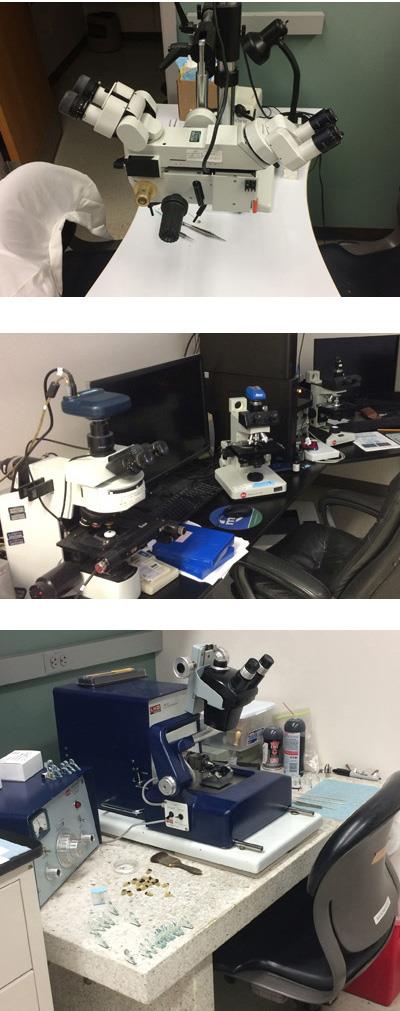
Histology
For tissue processing, there is an immunohistochemistry station, automated tissue processor and prep center, and an electron microscopy tissue processor. For tissue sectioning, there are two ultramicrotomes for thin sectioning of samples and a cryostat for frozen sections. There are also 2 glass-cutters for ultramicrotome blade construction. Additionally, there is a CLEMEX Vision PE imaging station suited for quantitative histology (histomorphometry).